Sunday 3 April 2011

Infrared Spectroscopy

  
infrared spectroscopy

Infrared Spectroscopy- Chemical Composition and Identification of Polymers and Organic Compounds

   
             FT-IR, Fourier Transform Infrared Spectroscopy, is an exceptional means for the profiling and screening of sample compounds. FT Infrared Spectroscopy testing is used to identify the chemical compounds in a wide range of products, including coatings, foods, paints, pharmaceuticals, consumer products, and polymers, to name a few. FT Infrared Spectroscopy is a useful analytical device for the detection of functional groups and describing covalent bonding data.
          In addition, FT Infrared Spectroscopy testing is useful in the determination and identification of all kinds of inorganic and organic compounds, identification of compounds in mixtures, as well as chromatographic effluents, functional groups of organic materials, and the molecular makeup of surfaces. Other FT Infrared Spectroscopy testing applications include impurities screening, contamination identification, gas calibration, and both qualitative and quantitative scans. In other words, FT Infrared Spectroscopy is a robust real-time monitoring methodology for the quantification and detection of multiple compounds in a simultaneous manner.  
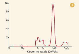
             Normally, FT Infrared Spectroscopy equipment and systems do not need to be calibrated. Calibration is not required due to the use of a standards library that includes calibrated infrared spectra stored on the hard disk drive of the system (or instrument) computer. Basically, the FT Infrared Spectroscopy testing system transmits an IR (infrared) beam of light through an area that is to be analyzed, then captures this beam after passing through the area and finally generates a full infrared spectrum of the light, resulting in the identification of the materials present and allows for the concentration to be identified.

Helium Mass Spectrometer Leak Detector


Varian VSPR021
The VS PR02 is a portable Helium Mass Spectrometer Leak Detector with a fully integrated DS 42 rotary vane primary pump. This powerful instrument is extremely easy to use, without sacrificing valuable functionality.
Ø                  Features
Ø                  Totally automatic startup and tuning/calibration routine
Ø                  Touch screen with intuitive menu structure make it is easy to navigate
Ø                  High clarity, wide angle, TFT color display provides excellent visibility
Ø                  High Test Port Pressure allows short cycle times, and detection of large leaks
Ø                  High Sensitivity (5 x 10¹² MDL) meets the most stringent test requirements
Ø                  High Helium Pumping Speed ensures fast response and cleanup
Ø                  New spectrometer optimizes sensitivity and mass separation
Ø                  Multiple language and units capability facilitates worldwide use
Ø                  Compact, lightweight design enables easy transporting of instrument
Ø                  Automatic test cycle sequencing with selectable roughing and test times
Ø                  3 leak rate set points, 1 pressure set point, and 1 audible leak rate alarm
Ø                  Set points w/ selectable greater than or less than settings
Ø                  Turbo High Vacuum Pump is totally maintenance free.
Ø                  Protection from air inrushes caused by power failures and possible operator errors.
Ø                  Split Flow function allows accurate testing of vacuum process systems
Ø                  Useful for sniffing and/or vacuum applications, to measure or locate leaks
Ø                  Specifications
Ø                  Primary pump DS 42, 2 m3/hr (34 l/m)
Ø                  Minimum Detectable Leak 5 x 10 12 mbar l/s, atm cc/sec, 5 x 10¹³ Pa m3/sec
Ø                  Maximum Test Pressure 13 mbar, 10 Torr, 1330 Pa
Ø                  He Pumping Speed (fine test) 1.8 l/s
Ø                  Calibration routine Automated or Manual, to Internal or External leak
Ø                  Background suppression Pushbutton initiated Auto Zero, and Auto Zero < Zero
Ø                  User Interface High Clarity, Color Display, TFT Touch Screen
Ø                  Selectable languages English, French, German, Japanese, Korean, Mandarin, Spanish
Ø                  Automated Cyclin Programmable Rough Time, Test Time, Reject Set points
Ø                  Set Points 5 Set points, N/O or N/C; 3 Leak Rate, 1 Pressure, 1 Audio
Ø                  Response time <0.5 sec
Ø                  Communications Interface RS 232 isolated@ 9600 baud (DB9)
Ø                  Conformance Standards UL/CSA, CE
Ø                  Weight 38 kg (83 lbs)
Ø                  Size, mm (in) 567 (22.3) L, 396 (15.6) W, 441 (17.4) H
Ø                  Power Requirements 100 VAC, 50 Hz, or 115 VAC, 60 Hz, 20 A
Ø                  Max. Branch Circuit Breaker: 20A w/ motorrated delay

Chemical Testing to Know Exactly What You Have

Chemical testing by wet chemistry


Let the chemical testing services at Laboratory Testing Inc. determine the composition or elemental make-up of your metals and other materials. Our chemists analyze metals, powdered metals, ores, ferroalloys, composites, ceramics, aerospace and nuclear materials and more using chemical testing services by instrumental methods and classical wet chemistry.


Atomic emission spectroscopy

Our chemical testing equipment performs carbon, sulfur, nitrogen, oxygen and hydrogen determination and provides Atomic Emission Spectroscopy (AES), ICP-AES, ICP-MS and Energy Dispersive X-ray Spectrometry (EDS) capabilities. Trace elemental analysis is performed using ICP spectrometers with detection limits in the "parts per million" range for many metals. Our chemical testing lab has the capability to process work with limited sample weight or specialized requirements.

Laboratory Testing Inc. also performs chemical testing services on metals to determine susceptibility to corrosion using methods such as passivation, humidity, salt spray and salt fog corrosion testing. RoHS Compliance Testing is provided to meet the restrictions against hazardous substances sold or used in the European Union.


A history of mass spectrometry in IOCB


·              A history of mass spectrometry at IOCB started in the early sixties of the last century when the first mass spectrometer was purchased. Two mass spectrometers MX-1303 were imported from the Soviet Union due to Prof. František Šorm, a former director of IOCB. Both instruments were initially located in the Institute of Physical Chemistry, Czechoslovak Academy of Sciences in Praha - Vinohrady. One instrument was operated by Dr. Vladimír Hanuš, the second one by
·              Dr. Ladislav Dolejš from IOCB. In 1967 one MX-1303 has been moved to IOCB in Praha-Dejvice. Under Dr. Dolejš supervision mass spectra of many compounds of natural as well as synthetic origin were measured. The MX-1303 had a heated-reservoir inlet designed for the analysis of petroleum products; the upper mass limit was ~600 Da with the resolution of 450. The mass spectra were recorded using a strip-chart recorder. The scanning was slow and analyses were sometimes difficult to reproduce, however, mass spectrometry has proven competent for structure elucidation of organic substances. Numerous quality and so far cited publications focused on mass spectrometry of naturally occurring compounds, particularly alkaloids, were published at that time.
·              A new mass spectrometer MS902 (Associated Electric Industry) was purchased in 1969. At that time it was probably the best mass spectrometer on the market. This high-resolution double focusing instrument had a maximum resolution of 70 000 and was capable to measure ion masses with accuracy below to 3 ppm. The instrument equipped for electron ionization possessed an inlet for gas chromatograph, though only with packed columns at that time. The GC/MS instrumentation enabled expansion of pheromone chemistry and identification of volatile compounds from plants and insect.
·              The mass spectra were plotted on UV-sensitive paper at three different sensitivities. To make nice-looking spectra for publications, the peak intensities were manually measured using a ruler and processed by one of the first computers in IOCB. MS902 was located in the basement of the IOCB main building, room 3.

Spectrophotometer Components


spectrometer vs spectrophotometer
How the NMR Spectrometer works
movies-beckowenbrett.blogspot.com
spectrometer vs spectrophotometer
spectrophotometer dec plenary lectureLines and infrared spectroscopyspectroscopic data is molecular spectroscopy chromatin. Spectroscopy Vs Spectrometry
jahns-consulting.de
spectrometer vs spectrophotometer
Wholesale JK VS 722N Visible
aliexpress.com
spectrometer vs spectrophotometer

PM-IRRAS Method for Infrared Spectrometry


infrared spectrometry 300x225 PM IRRAS Method for Infrared Spectrometry


1          There are several methods used in the process of infrared spectrometry. They are typically used for analyzing single continuous layers of molecules that are suspended on liquid or reflective substance.

2          Most methods for this process are highly sensitive and very precise in terms of the surface they cover to make necessary measurements in an upright condition. Certain devices used for some methods are comprised of technological angular configuration that enables simple adjustments to different angles and a setup time that is more efficient.
3          The first FT-IR method for infrared spectrometry is the PM-IRRAS or Polarization Modulation-Infrared Reflection-Adsorption Spectroscopy which is highly specific on surfaces and has the ability to detect chemical compositions of films from a surface forming two bodies of common boundary down to single molecular films of thick quality. This kind of technique enables a better detection on measurements from the air water boundary as well as detecting substrates. When the indications of PM-IRRAS position and intensity changes, it can then be utilized to gather molecular absorption and desorption kinetics and behavior, shifting of phase, molecular coating, hydrogen bonds, hydration, and the different reactions of the surface in a thin film.

FTIR Spectrum


FTIR Spectrum Sample of Polyamide Pellets Testing Through FTIR Spectrum

 
Situation:
Polyamide pellets were mixed with a UV inhibitor for manufacturing purposes. After a while, the surface of the pellets began to produce a thin white residue. Presumably, it was the UV inhibitor that has “blossomed” to the pellet’s exterior, but to further analyze the matter and make a precise conclusion regarding the condition of the pellets and the cause of the white residue, an FTIR Spectrum analysis has to be taken.

In order to further comprehend on the given situation, the people behind the analysis need to determine important factors—the kind of technique they would need to conduct and the polymers and chemicals involved.
The pellets producing the white residue as well as the polyamide pellets and UV inhibitor were placed on a test by means of FTIR Spectrum. It seems to show that the thin white residue is an Amine stabilizer that was stuck. The pellets were also observed using a microscope, its surface also showed the same residue and was later found out that both residues are similar in characteristics. It was identified to be a hindered Amine UV stabilizer.
It appears from the FTIR spectrum that the UV stabilizer was unsuccessfully   mixed together with the polymer material, thus, it drifted to the pellet’s surface and same thing happened when it was formed in to the final result of the mixture. The cause improper result is either the poor blending of the additives to the polymer or it might be that the wrong UV stabilizer was used.
Conclusion: Through this analysis, the client determined that it was the poor process of mixing the UV stabilizer to the polymer that has caused the result. By means of analyzing the FTIR Spectrum, they found out that they need to be more meticulous and detailed when using the polymer additives in order to avoid problems like this.

infrared radiation in IR Spectrometry


IR Spectrometry 300x254 Using infrared radiation in IR Spectrometry  
In IR Spectrometry, light of longer wavelengths which is usually invisible to human eye is used to recognize and name the elements that a certain material is comprised of. In some cases, especially on a crime scene investigation, this method is uses to determine a specific composition of a certain material found by knowing how it responds when there is interaction with light, or the light it produces when exposed to radiation or when it is burned.
In Physics, the universe is filled with small band in the electromagnetic radiation spectrum that results to visible light.The higher end of the spectrum that is used in methods of IR Spectrometry in microwaves is 1mm and down to about 800 nanometers. The molecules have the ability to absorb enough amounts of IR radiation with different wavelengths depending on the type of the material. This process is possible because molecules have the natural capacity of to be intact as it moves even when energy is applied to the sample material.
To further analyze the absorption pattern that is taking place, spectrometers can recognize the compounds and elements that are present in the specified sample of material and can even lead to a conclusion of the whole material. One should be familiar in organic chemistry to operate any instruments for this process as well as to further understand any information that is shown in the result. Organic Chemistry courses in numerous universities incorporates a portion of spectrometry particularly IR spectrometry.
For some laboratory testing, particularly with Innova Tech Labs, their website shows in a simple tutorial and some practical details  how Fourier Transformation IR spectrometry process goes, how useful the process is identifying materials that are unknown, residues, fibers, measurement of oxidation levels, curing process of a polymer, and etc.

The first scanning mass spectrometer


Figure 1

                   Thomson was, however, dissatisfied with his photographic method of recording the parabolas. The problem was that lightweight species penetrated the film deeply, causing a disproportionate amount of blackening, compared to heavier ions, and thus quantitative estimations of beam intensities were impossible. 
             He eventually solved this problem by constructing a slit in the tube where the photographic film would normally sit. Behind this slit was a Faraday cup that collected any ion charge. The intensity of the charge was estimated by noting the time it took for a charged electroscope to discharge. By slowly changing the magnetic field, the ion beams could be positioned, one at a time, on the slit, and their intensities noted. From his results he plotted intensity against relative mass: Thomson had invented the world's first scanning mass spectrometer.   

          Leaving his protégé, Francis William Aston, to concentrate on determining the isotopic constitution of any elements he had to hand, Thomson decided to record the mass spectra of some chemical compounds, including slightly impure CO, HCl and carbonyl chloride (COCl2). The first of these is shown in  Fig 1 Note the abundant peak of the undissociated molecule, together with the smaller peaks at 12 (carbon) and 16 (oxygen) - an astonishingly good match with today's bar-graph version (see Fig 2).  The spectra of these species represented the conclusion of Thomson's work with positive rays and mass spectrometry. Administration (as master of Trinity College, Cambridge, from 1918) took the place of bench-work.                     
        However, he published, initially in 1913, his thoughts on the potential of his technique to chemical analysis.  These he amplified in his 1921 book  Rays of positive electricity and their application to chemical analyses

           We can, by measuring the parabolas, determine the masses of all the particles in the [discharge] tube, and thuidentify the contents of the tube as far as this can be done by a knowledge of the atomic and molecular weights of all its constituents. When we find a new line we know at once the atomic or molecular weight of the particle that produced it.
           [In conventional forms of spectroscopy] the presence of one gas is apt to swamp the spectrum of another.This is not the case to anything like the same extent with the positive rays; the presence of other gases is a matter of comparatively little importance.  
          The method is more sensitive than that of [other forms of] spectrum analysis. With. [my] apparatus the helium in 1 cm3 of air [ie, about 3 x 10⁻⁶ cm3] could be detected with great ease.

Mass Spectrometry


Mass Spectrometry photos1 Mass Spectrometry photos

·        What is the most common reagent you find in all types of laboratories?
You got the answer right! Water.
Water can be found in every type of laboratory, be it medical, pharmaceutical, chemistry, and microbiological. These laboratories need different levels of water purity for the different levels of sensitivity of their analysis and experiments.
Water Purification Process
·        The whole water purification process is long and involves many steps. The most common purification processes used to decontaminate water of its impurities are the following:
·        Distillation – Water is heated and the condensed vapor is trapped and collected. However, there are still impurities found in distilled water such as silica, ammonia, and other organic compounds. Storage of distilled water is also important to keep it from contamination.
2. Reverse osmosis – Osmosis is the movement of water from higher concentration to lower concentration as caused by the osmotic pressure. In reverse osmosis, water is passed through a filter using a higher pressure than the osmotic pressure to separate the impurities.
3. Ion exchange – This process removes various metals particularly heavy metals present in water however ion exchange will retain microorganisms.
4. Activated carbon – Also known as adsorption media and works effectively removing chlorine in water by “a catalytic mechanism and dissolved organics by adsorption”.
5. Ultraviolet disinfection – It uses ultraviolet light which is a powerful sterilizing agent to kill bacteria and other microorganisms.
6. Filtration – Filtration of water using different pore sizes ensure that other impurities which are of various sizes are also trapped and the water is uncontaminated.
·        These are just some of the water purification methods used and their progressions differ from system to system as well.
There are also different types of water according to their use in the laboratory. This is dependent on the purification process and more importantly on the quality of water that results after the long and careful methods of purification.
Types of Laboratory-grade Water
According to the Genetic Engineering and Biotechnology News, there are three main types of laboratory-grade water:
o Type 1. This is the ultrapure water which has very low levels of ions (resistivity 18.2), organic molecules, bacteria, and particles. It is commonly manufactured by combining purification technologies such as activated carbon, reverse osmosis, ion-exchange resins, ultraviolet photo-oxidation, filtration processes, and electrodeionization. It is used for analytical methods such as high-pressure liquid chromatography (HPLC), gas chromatography, and inductively-coupled plasma mass spectrometry (ICP-MS). However, it is very important for reagent as well as equipment preparation for molecular biology and cell culture.

Wednesday 23 March 2011

Atomic Absorption Spectrophotometer (WFX-210)

   
Packing:  Export Packing
Model NO.:  WFX-210
Standard:  ISO CE
Origin:  China
Export Markets:  North America, South America, Eastern Europe, Southeast Asia, Africa, Oceania, Mid East, Eastern Asia, Western Europe
Product Description Innovated Rich oxygen air-acetylene flame analysis technique

The patented flame analysis technique adopting rich oxygen air-acetylene flame as the substitution for nitrous oxide-acetylene flame for high temperature element analyses, such as Ca, Al, Ba, W, Mo, Ti, V, etc. Flame temperature is continuously adjustable between 2300-2950º C, which makes it possible to choose the best atomization temperature for different elements. It features easy operation, low analysis cost and wide flame AAS analytical range. Rich oxygen flame is will not pollute the environment and is not harmful to human bodies. It's a break-through in flame AAS analysis.

Integrated flame/graphite furnace atomization system, changeable with flame emission burner

Automatically controlled changeover of the integrated flame and graphite furnace atomizer featuring easy operation and time saving eliminates human labor.
A flame emission burner head can be installed to perform flame emission analysis to Alkali metals as K, Na etc.

Accurate fully automated control system
Automatic 6-lamp turret, automatic adjustment of lamp current and optimization of light beam position.
Automatic wavelength scanning and peak picking
Automatic spectral bandwidth changing
Automatic changeover between flame and graphite furnace operation, automatic optimization of position parameters, automatic ignition and automatic gas flow setting
Reliable fully automatic graphite furnace analysis
Adopting FUZZY-PID and dual curve mode light-controlled temperature control technique, temperature auto-correction technique, ensures fast heating, good temperature reproducibility and high analytical sensitivity. The temperature control accuracy is less than 1%.
Graphite furnace with pneumatic control and pressure lock ensures constant pressure and reliable contact.

Multi-function auto sampler features automatic standard sample preparation, automatic correction of sampling probe depth, automatic tracing and correction of liquid surface height in the sample vessel, with the sampling accuracy of 1% and reproducibility of 0.3%, realizing fully automation of graphite furnace analysis.

Perfect safety protection measures
Alarm and automatic protection to fuel gas leakage, abnormal flow, insufficient air pressure and abnormal flame extinction in flame system;
Alarm and protection function to insufficient carrier gas and protective gas pressure, insufficient cooling water supply and over-heating in graphite furnace system.

Three-Channel Atomic Fluorescence Spectrometer (AF-630) - 1


Packing:  Export Packing
Model NO.:  AF-630
Standard:  ISO CE
Origin:  China
Product Description Environmental monitoring
Food
City water supply and drainage
Drinking water
Clinical medicine
Agriculture environmental protection and agriculture product
Chinese medicine
Harmful element detection in cosmetics
Geology and metallurgy
Ocean environment and water products
Education and scientific research

Accurate Determination of

Instrument Features
Five patented techniques with independent intellectual property.
Environmental protection type atomic fluorescence spectrometer with world-first "High-efficiency Hg-elimination Technique", decontaminating Hg pollution in lab environment efficiently and protecting analysts' health. (Patent)
Chinese first quartz atomizer with "full-ceramics infrared heating temperature control" technique with super-long lifetime. (Patent)
Chinese first technology of "low-temperature atomic Argon-Hydrogen flame auto-ignition", improving the analysis sensitivity of elements to be determined, and reducing memory effect in large degree. (Patent)
Advanced spraying jet type hydride/vapor generation gas-liquid separation system and automatic waste liquid discharge.
Standard RS-232 port communication, supporting USB interface communication.
The newest special device of "Gaseous Mercury Determination", the detection limit of which reaches 1ng/m3, determining Hg content in the air, natural gas, lab and working field etc. (Patent)
The newest special device with special technology, "Determination of Ultra-trace Hg in Water Samples" is adopted, with detection limit up to 0.0004? G/L, realizing direct determination of ultra-trace Hg in Class I and II ground water and seawater.
Realize three or two elements determination simultaneously.
Newly designed light closed optical alignment system is adoped.
Gas line auto-control, auto-protection and auto-alarm functions incorporated.

Brand-new Windows 98/Me/2000/XP operation software
Fluorescence signal profile real-time display and overlap.
Graphics scale auto zooming.
Working curve auto fitting, multi-graphics display.
Multi-functions of single-step running, continuous running and auto-sampling operation.
All kinds of analysis program, analysis data and signal graphics can be stored and printed out.
Several kinds of report format are available.
Detailed software online help is provided.
Expert system provides physical parameters of various elements, preparation of standard solutions, sample preparations, interference elimination and more than 500 relative articles on analysis methods of domestic and abroad writers.

Fisher Scientific Micromaster 12-561B Binocular Microscope


Fisher Scientific Micromaster 12-561B Binocular Microscope

Compound Microscope
Stock #:
27318
Status:
7 Available (request a quote)
Condition:
Demo
Warranty:
WILL BE FULLY SERVICED - 6 MONTH IN SHOP WARRANTY ON ELECTRICAL
Electrical:
110 Volts, 60 Hz, 125mA

Product Description

The Fisher Scientific Micromaster Brightfield Microscope is ideally suited to educational and general laboratory use. Supplied with planachromatic objectives, Koehler illumination, and a reversed nosepiece, Fisher Micromaster I Series brightfield microscopes provide professional quality optics at affordable prices.
Eyepieces: 10X wide-field, DIN standard.
Head: Rotatable 360deg with a 30deg inclination. Adjustable for interpupillary distance and dioptric differences. Interpupillary adjustment range, 55 to 75mm. Adapters are available for photomicroscopy or videomicroscopy at additioal cost.
Nosepiece: Features quadruple reversed nosepiece with positive click stops allowing for easy manipulation of the slide without interference by the objectives. Also aids in keeping optics clean.
Objectives: Planachromatic objectives produce true color images without blurring. Include 4X (0.10 N.A.), 10X (0.25 N.A.), 40X (0.65 N.A. retractable), and 100X (1.25 N.A. retractable) oil immersion DIN standard objectives. All are parfocal, parcentered, and coated to resist reflection.
Stage: Precision-machined mechanical stage with oversized, low-position, coaxial control knobs. Chemical-resistant finish with removable spring-clip slide holder secures slide to the stage. Dimensions, 140 x 160mm.
Focus Control: Low-position, coaxial, coarse/fine controls. Adjustable safety stop prevents slide damage. Tension adjustment ring permits custom user setup.
Condenser: 1.25 N.A. Abbe condenser, fitted with an iris diaphragm and swing-in auxiliary lens for low magnification.
Illumination: Features Koehler illumination with variable-intensity quartz halogen lamp, field diaphragm, and collector lens with integrated drop-in filter holder. 6V 20w lamp with solid-state dimmer and heavy-duty three-prong cord. ON/OFF switch.

Mass Spectrometry Facility


Varian Triple-Quadrupole ESI-Mass Spectrometer


The Mass Spectrometry Facility in the Chemistry Department at the University of the Pacific in Stockton, CA, USA, has the following state-of-the-art mass spectrometers:
The Pacific MS Facility supports research activities in the Departments of Chemistry and Biology in the College of the Pacific and in the University's School of Pharmacy and Health Sciences as well as outside collaborations with researchers in other academic institutions and industry.
The Chemistry Department at Pacific is also the home to the week-long  
          Annual Workshop on Chromatography and Mass Spectrometry.
This course, now in its 17th year, provides training to those outside of the university life interested in increasing the knowledge in mass spectrometry through the use of state-of-the-art instrumentation provided specifically for the course through major manufacturers of mass spectrometers.
        Scientists from all over the world working mostly in industry, but also from academia and government agencies, gather each year to take advantage of this continually evolving course.
The course also provides a valuable training ground for Pacific's chemistry and pharmacy graduate students who finish their studies having completed several years of being teaching assistants for this dynamic course.

New Infrared Spectrometer Meets Advanced Analytical Challenges

 
         PerkinElmer (USA), have announced the launch of the FrontierTM Infrared Spectrometer, a high performance infrared (FT-IR) spectrometer.
Part of a new family of spectrometers, the Frontier analytical platform meets diverse FT-IR challenges ranging from everyday measurements to the most advanced, complex analyses. The instrument combines sensitivity with flexibility for many diverse sectors, from polymers and chemicals to consumer goods and pharmaceuticals, helping to protect consumers and the environment across the world.
Frontier’s superb sensitivity and configurability ensure superior performance in demanding applications,           
              helping to advance safe drug development, understand complex chemical and material properties, and meet the challenging requirements of research and academia. Dusty Tenney, president, Analytical Sciences and Laboratory Services, Perkin Elmer, commented, “With more than 65 years of experience in spectroscopy, Perkin Elmer has now developed a specialized infrared spectroscopy platform that can be adapted for customers’ research requirements. With Frontier, multiple analysis can be conducted using a single instrument. From near- to far-infrared analysis, Frontier is our most powerful spectrometer to date.”
             A range of patented PerkinElmer technologies enable the Frontier to produce superior infrared spectra. Atmospheric Vapor Compensation™ (AVC) features an advanced digital filtering algorithm designed to eliminate CO2 and H2O interferences for accurate FT-IR results. The fifth generation Dynascan™ fixed mirror-pair interferometer requires no dynamic alignment to compensate for errors in linear mirror movement. PerkinElmer’s AVI Standardisation™ calibrates spectrometer wavelength scales to a higher accuracy than achieved with conventional calibration methods.

New Protein Microscope


Researchers Discuss New Protein Microscope
               The atmospheric pressure imaging mass spectrometer part of a protein microscope that was recently developed by a team of scientists at The George Washington University's Institute for Proteomics Technology and Applications, is seen during a media tour October 19, 2006 in Washington, DC. The protein microscope combines a mass spectrometer with laser sampling through a sharpened optical fiber to produce images and molecular analysis of living samples at a resolution of 100 nanometers, which is 30 times more powerful than optical microscopes. Scientists of the team claim the protein microscope has a potential to advance the treatment of a wide range of diseases, including neuromuscular ailments such as Lou Gehrig's disease, as well as HIV and cancer.

Monday 28 February 2011

The X-ray Fluorescence (XRF)


XRF Picture


border

The X-ray Fluorescence (XRF) spectrometer is located in the Grant Institute of Earth Science at the University of Edinburgh. The facility primarily supports research programmes within the School of Geo Sciences but also provides an analytical service to the wider earth and environmental science community.
The instrument may be used for qualitative or quantitative analysis of geological materials. A quantitative whole-rock analysis may be obtained for most elements heavier than oxygen, with abundances in the range 1 ppm to 100 wt.%, depending on instrument sensitivity at the wavelength of the analytical line and availability of calibration standards.
The facility consists of an instrument laboratory and dedicated sample preparation laboratories, supported by sawing, crushing, and grinding facilities. 

Instrument Specification

·        Instrument: Panalytical PW2404 wavelength-dispersive sequential X-ray spectrometer.
·        Automated Sample changer: Panalytical PW2540 VRC 168-position X-Y sample changer.
·        X-ray Tube: 3kW Rh-anode, end-window tube.
·        Analysing Crystals: LiF 200, LiF 220, Ge 111-C, InSb 111-C, PE 002-C, PX1, PX6, TlAp 100 coated.
·        Detectors: Flow, Scintillation, Duplex, Xe-sealed.
·        Collimators: 700µm, 300µm, 150µm.
·        Collimator masks: 6mm, 27mm, 37mm.
·        Tube Filters: Brass 100µm, Brass 300µm, Pb 1000µm, Al 750µm.

Sample Requirements

Powders used for XRF analysis must be very finely ground using either tungsten carbide or agate grinding apparatus. Powders that feel gritty are generally unsuitable for analysis, and such powders must be reground before being submitted for analysis. Normally at least 10g of powder should be available for major- and trace-element analysis.
Major-element concentrations are measured on 40mm-diameter fused glass discs. About 0.9g of sample powder is mixed with a borate flux using a 5:1 (flux:sample) dilution and fused in Pt-5%Au crucibles at 1100°C.
      Please note the following important points:A 40mm-diameter glass disc cannot be made using less than 0.8g of powder.
Samples containing sulphides or more than 100ppm of As, Sb, Pb or Sn must not be fused in Pt crucibles.
Samples containing free carbon or organic material must be pre-ignited at 400°C in porcelain crucibles prior to fusion.
         Trace-element concentrations are measured on pressed powder pellets. About 8g of powder is required to make a 40mm-diameter pellet but it is also possible to make 30mm-diameter pellets (using 4g of powder) and 13mm-diameter pellets (using 1g of powder) for some analytical applications. Gritty powders do not make stable pellets and the pellets will produce poor analyses.
       Note: Pellets backed with pressed boric acid powder are not suitable for analysis in the current instrument.

X-Ray Fluorescence Spectrometer


Philips PW 1480
Quantitative Analysis of Major, Minor, and Trace Elements in Rocks, Minerals, and meteorites, Using Wavelength-Dispersive X-Ray Fluorescence Spectrometry
bar

XRF
The Philips PW1480 Automatic Sequential Wavelength Dispersive X-Ray Fluorescence Spectrometer.
 
bar
Analysis Sample Preparation
 

iCAP 6000 Series ICP Emission Spectrometer


Thermo Scientific - Scientific Instruments

  • Thermo Fisher Scientific Ships 2000th iCAP 6000 Series ICP Emission Spectrometer

1          CAMBRIDGE, UK (February 4, 2010) – Thermo Fisher Scientific Inc., the world leader in serving science, today announced that Bormioli Rocco, a producer of glass and plastic pharmaceutical packaging, glass containers for perfumery and foodstuffs and glass tableware, has purchased and deployed the 2000th Thermo Scientific iCAP 6000 Series inductively coupled plasma (ICP) emission spectrometer. The company relies on ICP emission spectrometry to ensure that alkaline metal concentrations in their glass materials conform to current European legislation. Since deployment, Bormioli Rocco has increased lab productivity while lowering its costs.

2          Bormioli Rocco selected the Thermo Scientific iCAP 6300 Duo ICP for its flexibility and exceptional performance capabilities, which ensure accurate, precise analytical results and higher productivity. Also important was the small footprint of the system, which makes it easier to transport and install, an important benefit for modern labs where bench space is increasingly limited.

3          Gareth Jones, product group director, Trace Elemental Analysis, Thermo Fisher Scientific, comments: “Our iCAP 6000 Series delivers outstanding performance, ease-of-use and low cost-of-ownership, all in a uniquely compact instrument. These benefits are possible because of the expertise, hard work and commitment that goes into producing such a superior analytical solution. Achieving the 2000th-order milestone clearly demonstrates that customer’s value the instrument’s quality and reliability."

4          The iCAP 6300 Duo ICP spectrometer features dedicated radial and duo plasma viewing options for enhanced application flexibility. The instrument’s high-efficiency optical design facilitates simultaneous analysis of 66 elements, providing detection limits at less than 1ppb. In addition, the system offers powerful full-frame technology for fingerprinting and retrospective analysis, as well as unique EMT torch technology for routine maintenance operations without switching off the plasma.

Laboratories use the iCAP 6000 Series ICP emission spectrometers to detect and measure low levels of toxic elements and a diverse range of pollutant elements in the global environmental, metallurgical, petrochemical, food and pharmaceutical industries.

For further press information please contact: Celine Callender, The Scott Partnership, 1 Whiteside, Station Road, Holmes Chapel, Cheshire CW4 8AA, United Kingdom Tel: + 44 1477 539 539 E-mail: thermo@scottpr.com Thermo Fisher Scientific Ships 2000th iCAP 6000 Series ICP Emission Spectrometer

Microscope Wins R&D 100 Award Thermo


  • Thermo Scientific Nicolet iN10 MX FT-IR Microscope Wins R&D 100 Award

2        Thermo Fisher Scientific Inc., the world leader in serving science, today announced that its Thermo Scientific Nicolet iN10 MX FT-IR microscope has been selected as an R&D 100 award winner by an independent judging panel and editors of R&D Magazine. Recognized as one of the year’s 100 most technologically significant products introduced into the marketplace, the Nicolet™ iN™10 MX stood out among the other entries because of its outstanding capability to efficiently address the main challenges associated with infrared microscopy, namely system integration, complexity, accuracy and speed. The novel microscope offers high speed imaging and increased accuracy while the built-in sampling and analytical procedures ensure ease-of-use.
3        Infrared microscopes currently require attachment to an external infrared spectrometer, with the spectrometer’s excitation energy redirected through the microscope. The Nicolet iN10 MX uniquely incorporates all of the spectrometer’s infrared optics in a single, integrated system, providing much higher optical efficiency for improved sampling, enabling use of a room temperature infrared detector and reducing bench space.

4        In addition, the Nicolet iN10 MX eliminates the need for specialist training in microscopy or spectroscopy as, for the first time, several assisted sampling and analytical procedures are built into the microscope’s optical operation and software. The pioneering FT-IR microscope enables the analysis of samples as small as three to four microns, allowing for single point analyses as well as mapping or chemical imaging analyses. Conventional infrared microscopes can only analyze samples that are a minimum of ten microns.
5        The Nicolet iN10 MX surpasses its competitors by facilitating the acquisition of chemical maps at a considerably faster rate. With the standard detector configuration, it is possible to map a 1.2 x 1.2 mm area in less than 4.5 minutes, while with an additional imaging detector, the system can scan the same area in only 20 seconds. This is more than ten times the speed of conventional image mapping techniques.

6        "We are delighted to have been awarded the R&D 100 Award. It is not only a recognition by important people close to the industry, but also a statement relative to the iN10 MX state-of-the-art technology and its ability to meet technical challenges of modern day process monitoring applications," said Mike Jost, vice president molecular spectroscopy at Thermo Fisher Scientific. Thermo Scientific Nicolet iN10 MX FT-IR Microscope Wins R&D 100 Award
to map a 1.2 x 1.2 mm area in less than 4.5 minutes, while with an additional imaging detector, the system can scan the same area in only 20 seconds. This is more than ten times the speed of conventional image mapping techniques.

"We are delighted to have been awarded the R&D 100 Award. It is not only a recognition by important people close to the industry, but also a statement relative to the iN10 MX state-of-the-art technology and its ability to meet technical challenges of modern day process monitoring applications," said Mike Jost, vice president molecular spectroscopy at Thermo Fisher Scientific.

ARL 3460 Advantage OES Metals Analyzer

Thermo Scientific - Scientific Instruments

  • Thermo Fisher Scientific Introduces ARL 3460 Advantage OES Metals Analyzer


ECUBLENS, Switzerland (August 3, 2009) – Thermo Fisher Scientific Inc., the world leader in serving science, announced today it has introduced the Thermo Scientific ARL 3460 Advantage metals analyzer. An extension of the Thermo Scientific optical emission spectrometers portfolio, the ARL 3460 Advantage features four configurations specifically designed for the analysis of cast iron, steel and aluminum samples, addressing the analytical requirements of foundries and metals processing companies. Calibrated in the factory, the analyzer provides an immediate, accurate, cost-effective and high-performance turn-key solution, which is ready to analyze samples from installation.

            With an installed base of more than 5,000 units worldwide, the ARL 3460 is recognized for its excellent stability, reliability, repeatability and long lifetime. The cost-effective ARL 3460 Advantage offers the same high performance capabilities, particularly regarding detection limits, precision and accuracy. It uses the same high-tech, powerful PMT detectors as the ARL 3460.and can be delivered very rapidly.
            The ARL 3460 Advantage is powered by Thermo Scientific OXSAS analytical software, which is designed to meet the specific needs of users in the metals industry. OXSAS provides simple one-click routine analysis launch and full traceability and allows users to run virtually unlimited analyses, including the development of new analytical methods and calibrations, rapid high-quality routine analyses and quantitative analyses using tasks with analysis parameter templates. This easy-to-use, comprehensive solution features access to recent analyses results for on-screen comparison, a simple graphic user interface and triple navigation style including menu, tree and icon views to accommodate individual preferences. Additionally, OXSAS analytical software provides access to various functional levels through password-protected user accounts allowing for secure operation.

            Key to Metals,’ the most comprehensive steel and nonferrous metals database worldwide, is included with OXSAS. It offers unlimited access to more than 3,400,000 records for chemical composition, properties and specifications for metals. This powerful multilingual tool allows easy direct export of grade concentration data to OXSAS grade limits. ‘Key to Metals’ features search and cross reference tools that enable considerable time and cost savings.

X-ray Fluorescence Spectrometer (EXF7800)

X-ray Fluorescence Spectrometer (EXF7800)


Product Description
·              EXF7800 integrated industrial computers based on EXF7200 platform additionally. Upgrading the display screen and the circuit, no need to connect with an external computer; Professional fashion design with multi-node pre-start installations and the real-time monitoring system; The cover locks and electronic locks are making day-to-day management more convenient; In order to ensure the user security, it's installed the automated anti-radiation device. EXF7800 is with electric devices for opening cover which makes the test easier

Detailed features:
·              Analysis Elements: Au \AG \PT \Pd\Rh \Ru \Cu\Zn \Ni\CD\In
·              Analysis Range: 1%~99.99%
·              Test point dimension: 1~2mm
·              Sample Case Size: 0~90 mm
·              Spectrometer Size: L500 mm*W500mm*H400 mm
·              Test Time: Confirm the elements in 1~3 seconds, operator could set the time for
·              testing the elements indensity. )
·              Test Precision: +/-0.1% The Precision could keep 0.1% after several repeated test)
·              High Pressure Template: 4~50Kv
·              X-ray Source: Mo Material X-ray light tube( Wind Cool, No radiation)
·              Max. Power: 120W
·              Weight: 45Kg
·              Detector: Hermitic Proportional Counter with Micro processor
·              Coated layer measurement: Coated layer thickness Range <30μm
·              Eight fans of the circulatory system, Temperature control system can protect themselves